Diagnosis
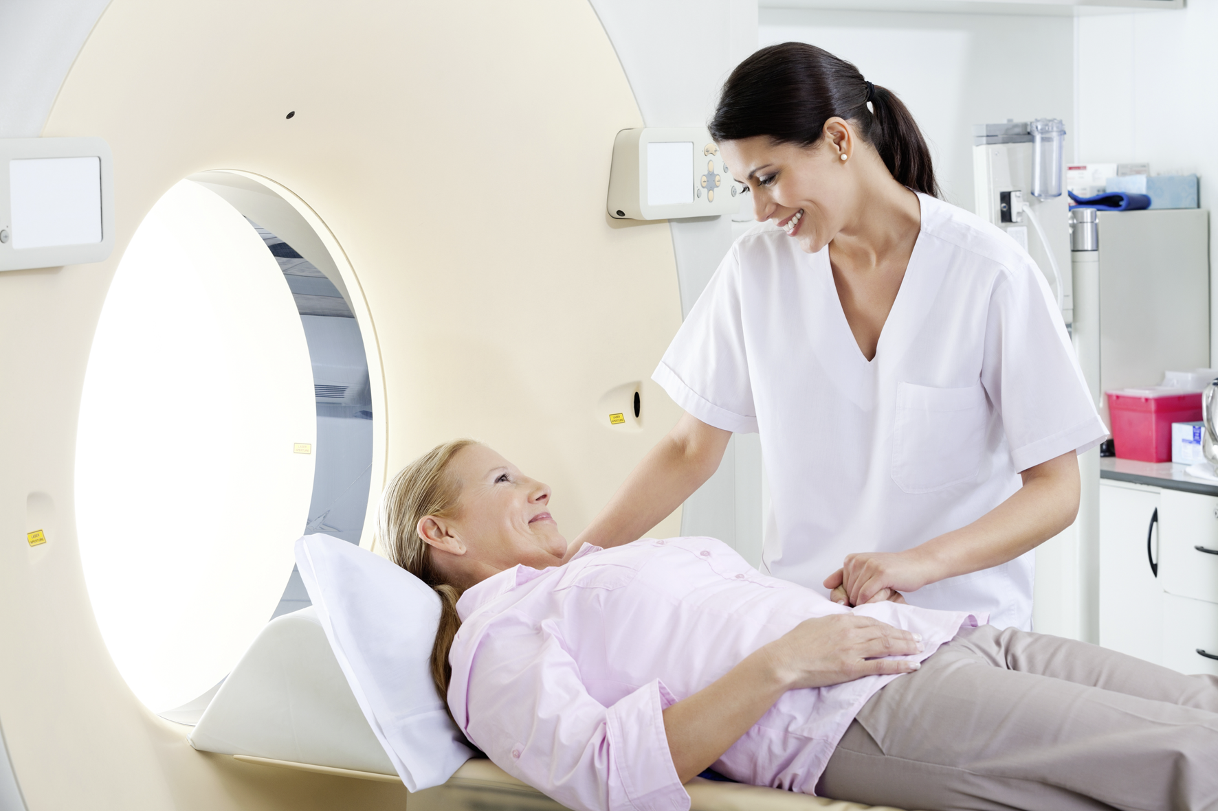
The first step to diagnosis is the evaluation of the patient’s medical history and a thorough clinical examination. However, when a swelling, a lump or pain is present, imaging tests are important to assist with diagnosis and staging. Imaging processes create detailed images of limb tissues and internal organs.
There are different types of medical imaging methods:
- Bone x-rays determine any damage to the bone, new bone growth or fractures
- Magnetic resonance imaging (MRI): MRI is the standard imaging modality. It creates images of the affected bone and also tissues surrounding it allowing doctors to assess whether the disease has spread to other locations in the body. MRI is especially helpful in imaging of the extremities, pelvis, and trunk.
- Computed tomography (CT) can be used to make bone damage as well as lesions in other parts of the body (e.g. lungs or other organs) visible.
- Positron emission tomography (PET) is also an effective imaging modality to determine whether the cancer has spread to other organs. It can also be used to evaluate the effect of a therapy on the tumour.
- Radionuclide bone scans help show if a cancer has spread to other bones. It can find metastases earlier than regular x-rays. Bone scans also can show how much damage the primary cancer has caused in the bone.
The results of the imaging process are important for further therapy planning.
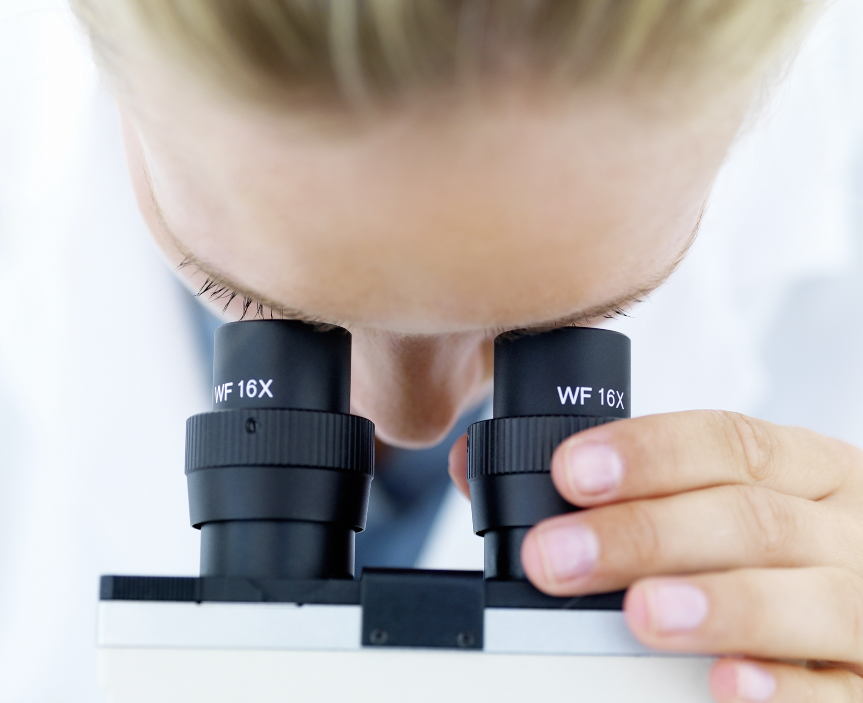 However, only a biopsy – removal of a sample of tumour tissue – can provide a definite diagnosis. A pathologist analyzes the tissue sample under the microscope and tests it in the laboratory. This so-called histopathological examination can determine if the tumour is a bone sarcoma and if so, what type. There are different types of biopsies: needle and incisional biopsy, which are chosen according the location of the tumour. During a needle biopsy, a small hole is made in the bone, and a tissue sample is removed from the tumour with a needle-like instrument. During an incisional biopsy, the tissue sample is removed after a small cut is made in the tumour. It is important that the needle or incisional pathway is excised during the main surgery.
Additional blood tests can help find bone cancer and might reveal further information about the specific type. In patients with osteosarcoma or Ewing sarcoma, certain enzyme levels (alkaline phosphatase or lactate dehydrogenase) might be elevated in the blood.